Your health in motion: How a vein ultrasound helps us decide the best treatment for you
Most vein problems start deeper than what you see on your skin. In many cases, chronic venous insufficiency (CVI) causes the blood pooling that leads to spider veins, varicose veins, swelling, and nighttime cramps. That’s why it’s important to visit a vein clinic with certified vein doctors who can diagnose the root cause before recommending a vein treatment.
A quick visual look isn’t enough; you need a precise map of how blood flows in your legs.
At Vein Treatment Clinic in Bethesda, we use ultrasound to thoroughly evaluate circulation in a comfortable setting. Our office is located at 6903 Rockledge Dr Suite 470, Bethesda, MD 20817, near Westfield Montgomery and Walter Johnson High School, with easy access from the Red Line (Medical Center Station and Bethesda Circulator).
What is a vein scan?
A vein scan is a noninvasive test that uses sound waves to create real-time images of your veins. In everyday terms, it’s an ultrasound that shows your vascular anatomy and how blood moves through it.
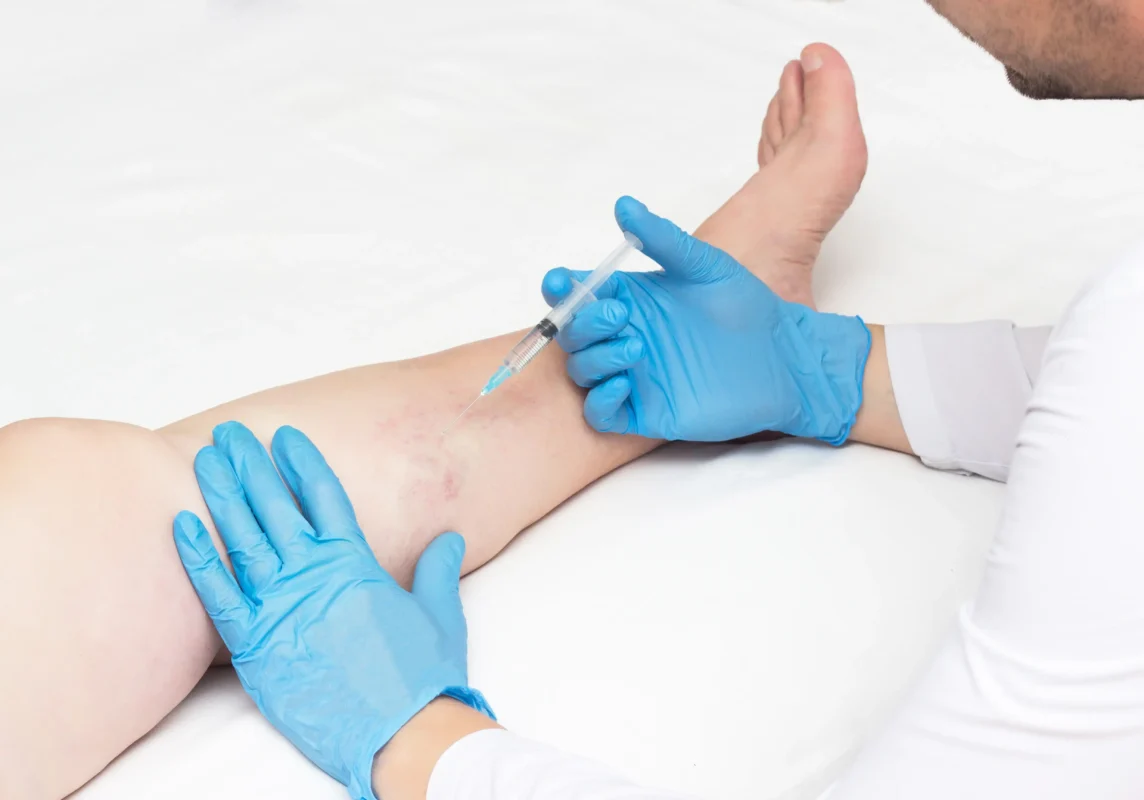
When you hear patients mention vein scan in Bethesda, they’re usually talking about this quick exam. There’s no radiation, no needles, and no downtime. It’s the gold standard we use to plan precise, minimally invasive treatments such as radiofrequency ablation or sclerotherapy, only when appropriate for your unique anatomy.
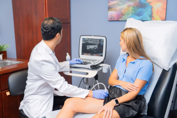
Dr. Kamran Saraf of Vein Treatment Clinic performing a vein ultrasound
Why vein Imaging matters before treatment?
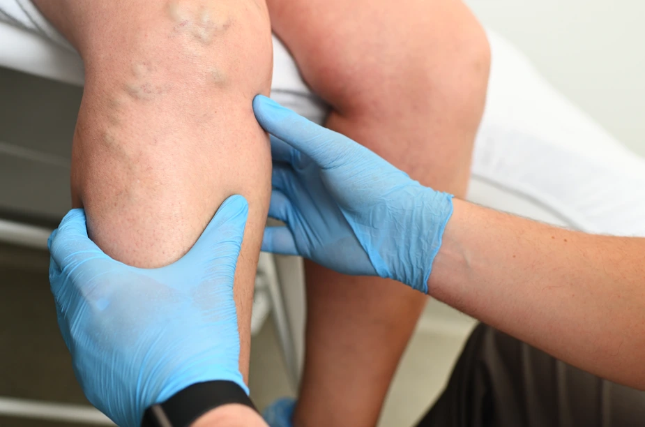
Chronic venous insufficiency happens when tiny one-way valves inside your leg veins don’t close properly. Normally, your calf muscles pump blood upward, and the valves in your veins prevent backflow, keeping blood moving toward the heart.
With CVI, your vein valves leak, allowing blood to reflux downward and pool in the superficial veins.
Over time, this pressure stretches the vein walls, leading to spider and varicose veins visible on the surface. Because the problem begins with deeper reflux, proper venous imaging is needed to confirm where the leak starts (often the great saphenous vein, small saphenous vein, or perforators).
How does early, accurate imaging benefit patients?
By mapping blood flow in real time, we can pinpoint exactly where valves need support and identify issues before they become visible. It’s the difference between guessing and having a personalized roadmap to recovery.
With accurate vein imaging, your vein doctor can:
- Identify the source of reflux, allowing for more targeted treatment
- Detect early valve dysfunction before symptoms progress
- Assess skin and tissue changes that may indicate advancing CVI
- Evaluate risk factors for venous ulcers
- Create a treatment plan based on your unique anatomy
Every patient’s journey is unique. That is exactly why we prioritize imaging to fully understand your specific circulation before creating a treatment plan tailored just for you
What to expect during your vein ultrasound?
One of the most common questions we hear: What actually happens when I book an appointment at the Vein Treatment Clinic? The answer is simpler than most people expect.
Your first visit will include a conversation about your health history, a physical examination of your legs, and a painless ultrasound that shows us exactly what’s happening beneath the surface. By the time you leave, you’ll have real answers, not guesses, and a clear path forward.
1. A thorough medical history
Your evaluation begins with a detailed conversation about your vascular history. Our vein specialist will focus on your overall well-being by listening to your specific symptoms. Some patients usually report some symptoms such as persistent aching, heaviness, nighttime cramps, or restless legs. This clinical intake ensures we understand the full story of your circulation before the imaging even starts.
2. Physical examination
Next, we perform a focused physical exam to identify the signs of venous health. We look for indicators like visible veins, swelling (edema), or changes in skin texture and color. This step allows our vein doctor to prioritize the most important areas during your ultrasound, ensuring your results are as accurate as possible.
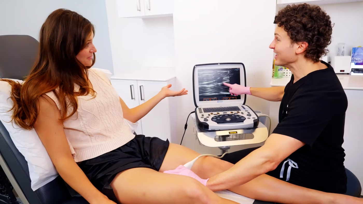
3. Ultrasound exam
Using advanced technology, we map your blood flow in real time to confirm healthy venous function and identify exactly where blood may be pooling in the legs. This allows us to treat the root cause of your symptoms, ensuring your path to recovery is based on clear, visual evidence.
Ready to start your journey? Book your appointment at the Vein Treatment Clinic and take the first step toward happier, healthier legs.
You’ll be welcomed by our front desk team and complete a brief medical intake. We will verify insurance for vein evaluations. If you need help clarifying the benefits of vein ultrasound in Bethesda, we’ll walk you through it. Contact us to verify your insurance details.

Stop guessing. Start healing.
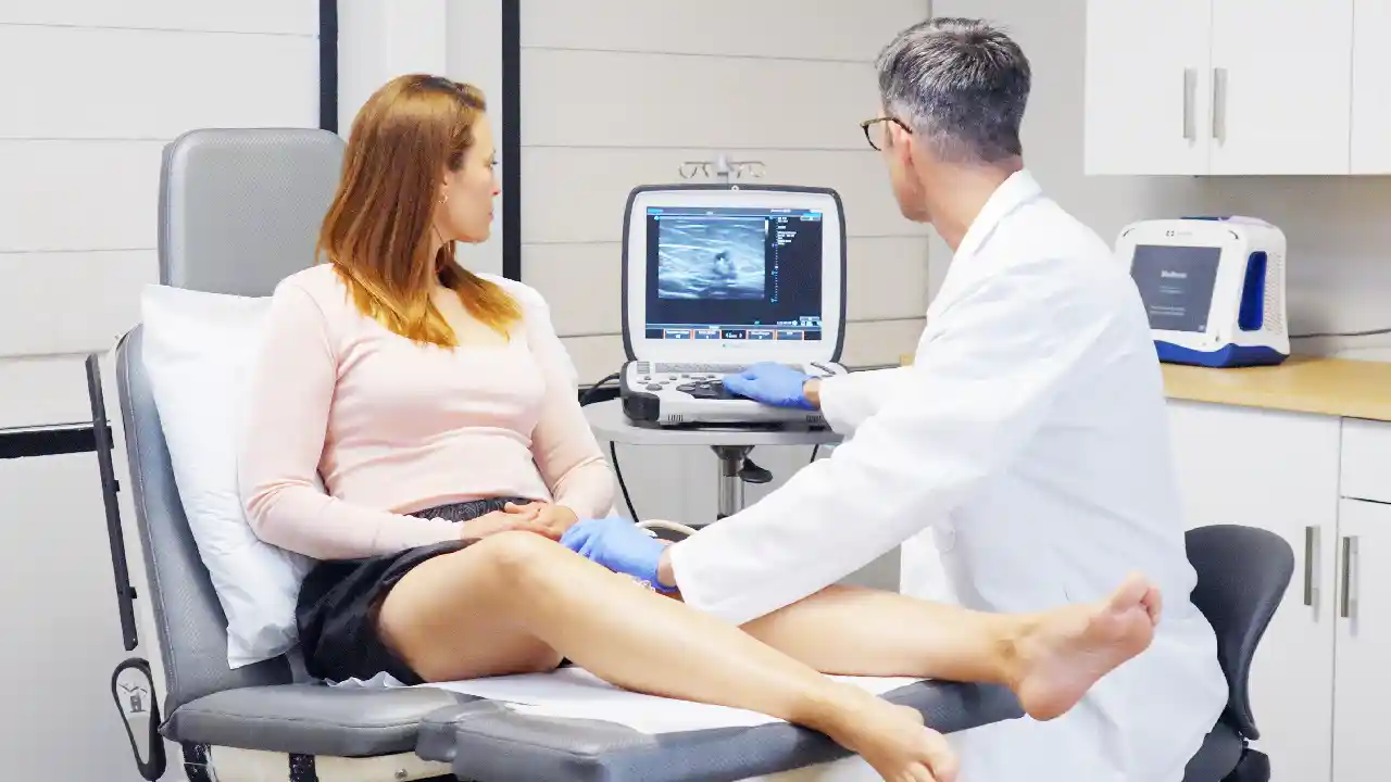
Why is a professional vein scan an important step in your recovery?
When you visit Dr. Kamran Saraf, your treatment isn’t based on a quick look; it’s built on a detailed map of your unique circulation. Using advanced ultrasound to confirm exactly where blood flow needs support, Dr. Saraf performs highly effective procedures with only local anesthesia and minimal downtime.
This evidence-based approach allows us to address the source of reflux first, so treatments like radiofrequency ablation or sclerotherapy can be tailored to your specific situation.
Meet the doctor behind your vein treatment
Want to know who’s reading your ultrasound and guiding your treatment?
Behind every diagnosis is a doctor who takes the time to get it right. Dr. Kamran Saraf leads our Bethesda vein center.
Dr. Kamran Saraf is the board-certified vein specialist leading our Bethesda clinic. His training spans Georgetown University, the University of Miami, Texas Medical Center, and the University of Pittsburgh and he’s earned the “Patient’s Choice Award” every year he’s been in practice.
But credentials only tell part of the story. Patients describe him as someone who never rushes, explains every step, and actually listens. He speaks English and Farsi.
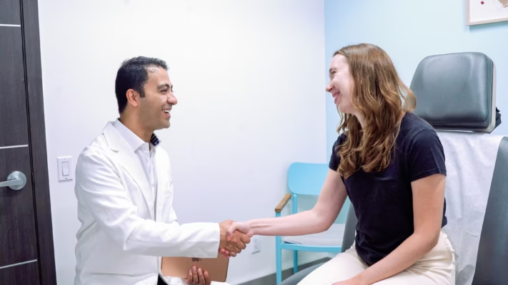
Meet Dr. Saraf from Vein Treatment Clinic
Take the first step toward lighter, healthier legs
If you are noticing spider veins, swelling, or nighttime cramps, a professional ultrasound is the best way to understand the “why” behind your symptoms.
At Vein Treatment Clinic Bethesda, our advanced imaging precisely maps your circulation and provides a clear roadmap for your recovery. Contact us today to schedule your consultation and get the answers you deserve.
Frequently Asked Questions
Is a vein ultrasound painful?
Not at all. A vein ultrasound is a completely non-invasive and comfortable experience. You will only feel a small amount of warm gel and light, soothing pressure as the probe moves along your skin. Because there are no needles or incisions, you can walk out of our clinic and return to your normal routine immediately.
Why is an ultrasound better than a visual exam?
Surface appearance is only one small part of the story. A visual exam can’t see what’s happening beneath the skin. Our ultrasound technology maps “hidden” flow patterns to ensure your treatment addresses the actual root cause, not just the visible symptoms.
How long does a vein scan take?
Most diagnostic scans at our Bethesda vein clinic take about 30 to 45 minutes. This allows us to be thorough and precise. The duration may vary slightly depending on whether we are assessing one or both legs to ensure we have a complete picture of your vascular health.
Do I need to prepare before my appointment?
There is no special preparation required. You can eat, drink, and take your regular medications as usual. We simply recommend wearing comfortable, loose-fitting clothing and avoiding heavy lotions on your legs the day of your visit to ensure the clearest possible imaging.
Will insurance cover my vein imaging?
When symptoms such as pain, swelling, or skin changes are present, most insurance providers recognize the medical necessity for a diagnostic ultrasound. Our team is happy to help you verify your specific benefits and navigate the coverage process, so you can have peace of mind.